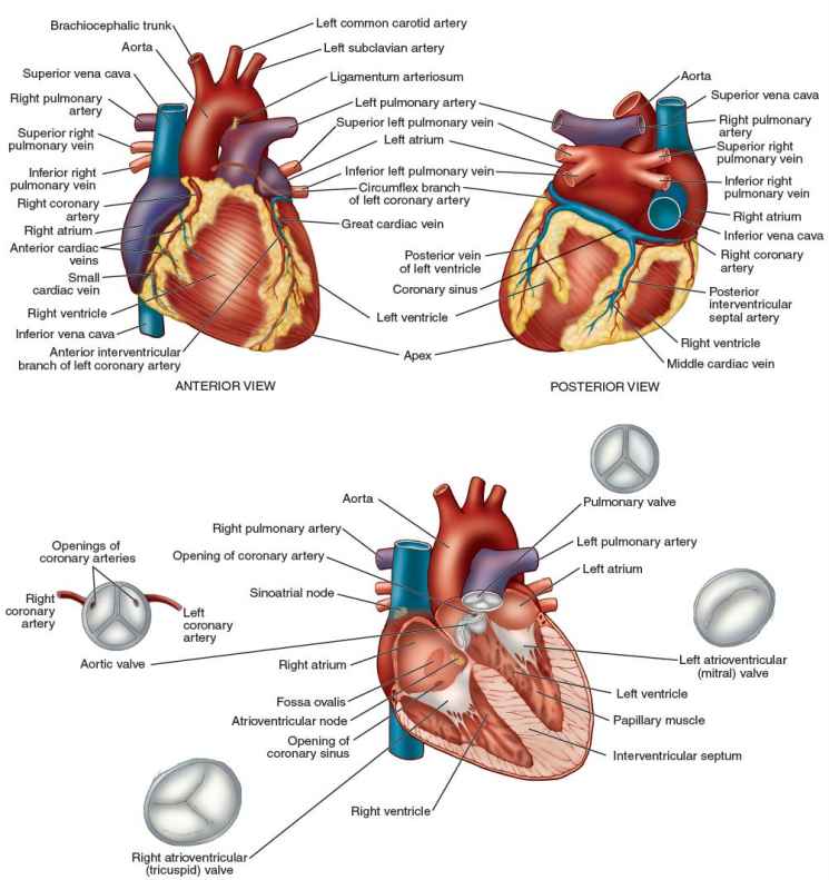
A hollow muscular organ that receives the blood from the veins and propels it into the arteries. In mammals it is divided by a musculomembranous septum into two halves—right or venous and left or arterial—each of which consists of a receiving chamber (atrium) and an ejecting chamber (ventricle). SYN: cor [TA], coeur. [A.S. heorte]
- armor h. extensive to complete calcification (rarely ossification) of the pericardium usually producing constrictive pericarditis.
- armored h. calcareous deposits in the pericardium due to subacute or chronic pericarditis. SYN: panzerherz.
- artificial h. a mechanical pump used to replace the function of a damaged h., either temporarily or as a permanent prosthesis.
- athlete's h. a more or less loose designation for cardiac findings in healthy athletes that would be or could be abnormal in patients with disease, including atrioventricular blocks, left ventricular hypertrophy and, sometimes, benign arrhythmias and atrioventricular blocks.
- beer h. SYN: alcoholic cardiomyopathy.
- beriberi h. h. disease due to thiamine deficiency that may be epidemic or sporadic as characterized by cardiac metabolic damage and myocardial failure, often of the “high output” type, with edema (except in “dry” shoshin beriberi) and polyneuritis. The term is derived from Singhalese, “I am unable.”
- bony h. the presence of extensive calcareous patches in the pericardium and walls of the h., some of which chronically develop bony changes.
- crisscross h. an anomaly in which the ventricular relationships are not as expected for the given atrioventricular connection.
- drop h. SYN: cardioptosia.
- fatty h. 1. fatty degeneration of the myocardium; 2. accumulation of adipose tissue on the external surface of the h. with occasional infiltration of fat between the muscle bundles of the h. wall. SYN: cor adiposum.
- frosted h. hyaloserositis involving the pericardium. SYN: icing h..
- globular h. SYN: round h..
- Holmes h. a variant of double inlet left ventricle where the ventricular-arterial connection is concordant and the right ventricle is rudimentary.
- horizontal h. description of the hearts electrical position; recognized in the electrocardiogram when the QRS in lead aVL resembles that in V6 and QRS in aVF resembles that in V1; also, loosely, when the electrical axis lies between −30° and +30°.
- hyperthyroid h. response of the h. to hyperthyroidism, essentially the result of sympathetic stimulation producing rapid h. rates and ultimately cardiac failure and atrial fibrillation if untreated.
- icing h. SYN: frosted h..
- intermediate h. loosely, description of the hearts electrical axis when this is directed at approximately between +30° and +60°. For cardiac position, recognized in the electrocardiogram when the QRS complexes in both lead aVL and aVF resemble that in V6.
- movable h. SYN: cor mobile.
- myxedema h. the enlarged h. associated with untreated severe hypothyroidism, often accompanied by pericardial effusion; rare in modern medicine.
- ox h. a very large h., due to chronic hypertension or, more often, to aortic valve disease, especially regurgitation. SYN: bucardia, cor bovinum.
- parchment h. a congenital or acquired condition in which there is thinning of the right ventricular myocardium. See Uhl anomaly. SYN: right ventricular hypoplasia.
- pendulous h. SYN: cor pendulum.
- pulmonary h. the right atrium and ventricle, receiving the venous blood and propelling it to the lungs. SEE ALSO: cor pulmonale.
- round h. abnormally smooth arcuate contours of the h. on imaging due either to disease of the ventricles or to a false cardiac appearance produced by excessive pericardial fluid. SYN: globular h..
- sabot h. SYN: coeur en sabot.
- semihorizontal h. loosely refers to the hearts electrical axis when this is directed at approximately 0°. As a cardiac electrical position, recognized in the electrocardiogram when the QRS complex in lead aVL resembles V6 while that in aVF is small algebraically or absolutely.
- semivertical h. loosely descriptive of the hearts electrical axis when this is directed at approximately +60°. As a cardiac electrical position, recognized in the electrocardiogram when the QRS complex in lead aVF resembles V6 while that in aVL is small algebraically or absolutely.
- systemic h. the left atrium and ventricle, receiving the aerated blood from the lungs and propelling it throughout the body.
- three-chambered h. congenital abnormality in which there may be a single atrium with two ventricles or a single ventricle with two atria. Rudimentary parts of the atrial and ventricular septa may be present but are incompetent to prevent a virtual single chamber in either case.
- tiger h. a fatty degenerated h. in which the fat is disposed in the form of broken stripes in the subendocardial myocardium.
- tobacco h. cardiac irritability marked by irregular action, palpitation, and sometimes pain, believed to occur as a result of the heavy use of tobacco.
- univentricular h. an anomaly in which all blood flows through one ventricle or in which the arterioventricular valves are committed to empty into only one chamber in the ventricular mass.
- vertical h. loosely descriptive of the hearts electrical axis when this is directed at approximately +90°. As a cardiac electrical position, recognized in the electrocardiogram when the QRS complex in lead aVL resembles V1 while that in aVF resembles V6.
- wooden-shoe h. SYN: coeur en sabot.
* * *
Healing and Early Afterload Reducing Therapy [study]; Health Education and Research Trial; Hyperlipidemia, Epidemiology, Atherosclerosis Risk-Factor Trial; Hypertension and Ambulatory Recording Venetia Study
* * *
heart 'härt n
1) a hollow muscular organ of vertebrate animals that by its rhythmic contraction acts as a force pump maintaining the circulation of the blood and that in the human adult is about five inches (13 centimeters) long and three and one half inches (9 centimeters) broad, is of conical form, is placed obliquely in the chest with the broad end upward and to the right and the apex opposite the interval between the cartilages of the fifth and sixth ribs on the left side, is enclosed in a serous pericardium, and consists as in other mammals and in birds of four chambers divided into an upper pair of rather thin-walled atria which receive blood from the veins and a lower pair of thick-walled ventricles into which the blood is forced and which in turn pump it into the arteries
2) a structure in an invertebrate animal functionally analogous to the vertebrate heart
* * *
n.
a hollow muscular cone-shaped organ, lying between the lungs, with the pointed end (apex) directed downwards, forwards, and to the left. The heart is about the size of a closed fist. Its wall consists largely of cardiac muscle (myocardium), lined and surrounded by membranes (see endocardium, pericardium). It is divided by a septum into separate right and left halves, each of which is divided into an upper atrium and a lower ventricle. Deoxygenated blood from the vena cavae passes through the right atrium to the right ventricle. This contracts and pumps blood to the lungs via the pulmonary artery. The newly oxygenated blood returns to the left atrium via the pulmonary veins and passes through to the left ventricle. This forcefully contracts, pumping blood out to the body via the aorta. The direction of blood flow within the heart is controlled by valve.
* * *
(hahrt) [L. cor; Gr. kardia] the viscus of cardiac muscle that maintains the circulation of the blood. Called also cor [TA]. It is divided into four cavities—two atria and two ventricles. The left atrium receives oxygenated blood from the lungs. From there the blood passes to the left ventricle, which forces it via the aorta through the arteries to supply the tissues of the body. The right atrium receives the blood after it has passed through the tissues and given up much of its oxygen. The blood then passes to the right ventricle, and then to the lungs, to be oxygenated. The major valves are four in number: the left atrioventricular valve (mitral), between the left atrium and ventricle; the right atrioventricular valve (tricuspid), between the right atrium and ventricle; the aortic valve, at the orifice of the aorta; and the pulmonary valve, at the orifice of the pulmonary trunk. The heart tissue itself is nourished by the blood in the coronary arteries. See Plate 18.
 PLATE 18—STRUCTURES OF THE HEART
Medical dictionary. 2011.